


Two researchers from the University of Notre Dame receive recognition for publications and images from the Notre Dame Integrated Imaging Facility (NDIIF).


Two researchers from the University of Notre Dame are recieving recognition for publications and images from the Notre Dame Integrated Imaging Facility (NDIIF). Each year, the NDIIF provides imaging awards to recognize and honor researchers who use the facility equipment in within each of the four imaging cores; Optical Microscopy, Electron Microscopy, InVivo Imaging, and Histology.
“We are grateful to all of the users of the NDIIF. Our annual imaging awards provide us an important opportunity to recognize their discoveries,” said Bradley Smith, Emil T. Hofman Professor of Science in the Department of Chemistry and Biochemistry and director of the NDIIF. “This year’s awardees showcase the breadth and the beauty of the research taking place within the facility.”
Best Publication Image Award Winner
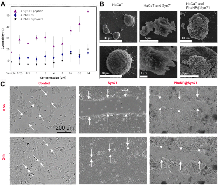
(A) Cytocompatibility of PhaNP@Syn71 on the HaCaT human keratinocyte cell line. PhaNP@Syn71 were cytocompatible at all concentrations tested similar to vehicle only control. Syn71 peptide was cytocompatible to 8 μM only. The average of four replicates for each sample was computed together with standard deviations. (B) SEM images of HaCaT cells incubated with PhaNP@Syn71. This indicated that the HaCaT cells integrity was not hampered by Syn71 or PhaNP@Syn71 at 8 μM concentration w.r.t Syn71. (C) In vitro scratch closure assay on HaCaT cells showed no cell migration in the presence of PhaNP@Syn71, while Syn71 peptide only presence, or no treatment group control allowed cell migration and scratch closure. This is because the PhaNP@Syn71 had a net negative charge, which will allow them to coat the positively charged tissue culture treated gap, as a consequence of which the surface charge of the gap becomes negative and repels cells migration. The free peptides are positively charged and do not hinder cell migration. 200 μm scale bar is applicable to all optical images. The images on the right represent histology samples. Images were taken a 10x magnification.
Kevin Armknecht, a distinguished undergraduate student, has received an award for best imaging publication! Kevin is a senior Science Pre-Professional Studies major with a minor in Compassionate Care in Medicine and Poverty Studies. He is currently conducting research in the Nallathamby Laboratory that looks at modular and scalable nanotechnology for imaging, sensing, and therapeutics. The winning image appeared in Nanoscale Advances in the article “Antimicrobial peptide-conjugated phage-mimicking nanoparticles exhibit potent bactericidal action against Streptococcus pyogenes in murine wound infection models”
Streptococcus pyogenes is a causative agent for strep throat, impetigo, and more invasive diseases. The main reason for the treatment failure of streptococcal infections is increased antibiotic resistance. In recent years, infectious diseases caused by pyogenic streptococci resistant to multiple antibiotics have been rising with a significant impact on public health and the veterinary industry. The development of antibiotic resistance and the resulting emergence of multidrug-resistant bacteria have become primary threats to the public health system, commonly leading to nosocomial infections. Many researchers have turned their focus to developing alternative classes of antibacterial agent based on various nanomaterials.
Kevin is a member of the Berthiaume Institute for Precision Health, led by Prakash Nallathamby, Associate Director of Research and Assistant Professor of Practice at The Notre Dame Berthiaume Institute for Precision Health (IPH) | Assistant Research Professor in the Bioengineering graduate program in Aerospace & Mechanical Engineering (concurrent).
Best Publication Image Award Winner
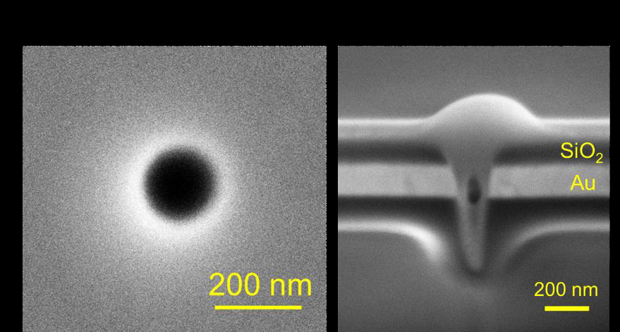
A) Magnified plan view SEM image of one nanopore. B) Cross-sectional SEM view of single pore showing 200 nm thick Au and 100 nm thick SiO2 hierarchical layers.
Jarek Metro, a graduate student in the Department of Chemistry under the direction of Professor Paul Bohn is receiving an award for best publication image for research published in Analytical Chemistry titled “Nanopore-Enabled Dark-Field Digital Sensing of Nanoparticles.”
The paper describes nanopore arrays as a platform for detecting and characterizing individual nanoparticles (NPs) in real time. Dark-field imaging of nanopores with dimensions smaller than the wavelength of light occurs under conditions where trans-illumination is blocked, while the scattered light propagates to the far-field, making it possible to identify nanopores. The intensity of scattering increases dramatically during insertion of AgNPs into empty nanopores, owing to their plasmonic properties. Thus, momentary occupation of a nanopore by a AgNP produces intensity transients that can be analyzed to reveal the following characteristics: (1) NP scattering intensity, which scales with the sixth power of the AgNP radius, shows a normal distribution arising from the heterogeneity in NP size, (2) the nanopore residence time of NPs, which was observed to be stochastic with no permselective effects, and (3) the frequency of AgNP capture events on a 21 × 21 nanopore array, which varies linearly with the concentration of the NPs, agreeing with the frequency calculated from theory. These results provide valuable insights into the dynamics of NP transport into and out of nanopores and highlight the potential of nanopore arrays as powerful, massively parallel tools for nanoparticle characterization and detection.
All awarded images were taken on NDIIF equipment and were taken by a Notre Dame faculty, staff, or student. Both of these accomplishments will be recognized at the 9th Annual Midwest Microscopy and Microanalysis Workshop on May 7, 2024.
The NDIIF at the University of Notre Dame provides an integrated suite of sophisticated microscopes and imaging stations that enable expert users to attack the most complex research problems as well as resident professional staff, including technicians and research specialists, to guide non-expert users. The facility is open to campus and external users. To learn more about the facility, please visit imaging.nd.edu.
Originally published by at imaging.nd.edu on March 20, 2024.
Latest Research
- Senator Todd Young and NSF Director Sethuraman Panchanathan visit Notre Dame to discuss critical investments in science and technologyOn Thursday, April 25, Sethuraman Panchanathan, director of U.S. National Science Foundation (NSF), joined U.S. Senator Todd Young in a visit to the campus of the University of Notre Dame. The pair met with faculty, students, and University leaders and discussed how research and innovation can drive…
- White House’s Jake Braun addresses statewide cybersecurity summit at Notre DameOn April 18, 2024, leaders in cybersecurity from government, industry, and academia gathered for the 2024 Indiana Statewide Cybersecurity Summit hosted by the University of Notre Dame in collaboration with co-sponsors Indiana University, Purdue University,…
- Hauenstein, Putman named as Simons FellowsTwo University of Notre Dame professors were named fellows of the Simons Foundation for 2024, which will allow them the opportunity to intensely focus on their research for up to a year. Jonathan…
- Literacy scholar Ernest Morrell elected to American Academy of Arts & SciencesErnest Morrell, the Coyle Professor of Literacy Education at the University of Notre Dame, has been elected to the American Academy of Arts and Sciences, one of the nation’s oldest learned societies and independent policy research centers. Morrell was one of the 250 members of the newest AAAS class announced today. Other notable names among the group include filmmaker George Clooney, Apple CEO Tim Cook, novelist Jhumpa Lahiri, and Pulitzer Prize-winning New York Times columnist and 1993 Notre Dame alumnus Carlos Lozada.
- Notre Dame faculty fight malaria resurgence in BangladeshBetween 2008 and 2020, districts across the country of Bangladesh saw a 93% reduction in malaria cases. Today, as the world reflects on the World…
- Anthropologist's research shows there’s no ‘one size fits all’ when it comes to addressing men’s health issues globallyAt a time when health resources are at a premium and need to be wisely allocated, health professionals must find points within men’s lives when it makes the most sense to intervene and advocate for preventive care for promoting better health outcomes. Life transitions such as marriage and fatherhood are often pivotal and crucial intervention points. But just like every man is different, health concerns across global communities differ as well. Research from the University of Notre Dame finds that not all life transitions produce the same health results, and not all men’s global health policies should look the same from one country to another.


